Ultrasound relies on the transmission and reception of sound waves above the range of human hearing. Ultrasound is used in hospitals and clinics to generate two- or three-dimensional images. To study a particular area of the body, a transducer sends out high-frequency ultrasonic waves and picks its echo.
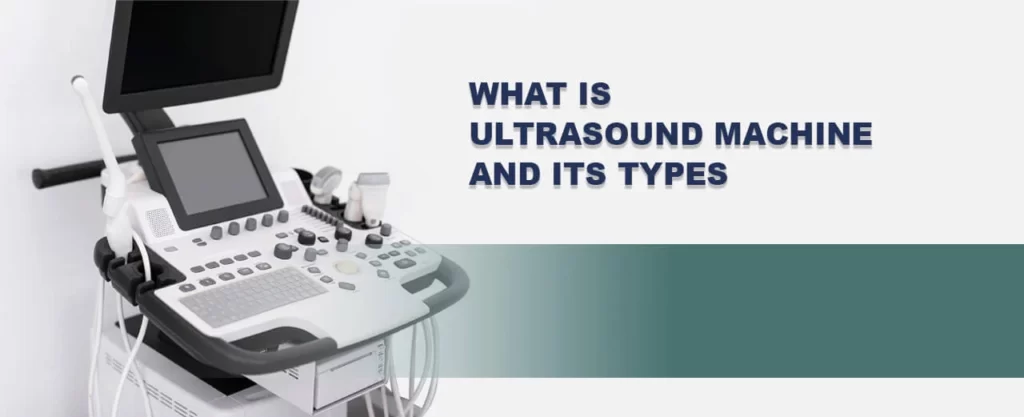
The transducer is the device that transmits and directs short waves with high-frequency, inaudible to humans, to the body interiors. It records any changes in sound, no matter how tiny they may be, by bouncing off sound waves through tissues, fluids, or organs. The transducer records an echo, and a computer transforms that echo into images that display on a screen. In an era of medical advancements, ultrasound scanning has gained significant recognition for impactful diagnosis and treatment of diseases. This blog will offer insight into ultrasound machines and their relevance in modern medicine. There is a considerable demand for ultrasound scanning devices among medical departments.
An Insight into Ultrasound Machine
The ultrasound scan procedure, also known as a sonogram, uses high-frequency sound waves to generate an image of a section of the inside of the body. We can employ ultrasound scans to track a fetus or inborn child, identify a medical issue, or direct a surgeon during certain surgeries.
The ultrasound machine emits high-frequency sound waves directed at the internal body structures under examination. The scanning machine records echoed or reflected sounds to generate an image displayed on a monitor. A small, handheld probe emits and receives sound waves.
To monitor the internal body, ultrasound imaging employs high-frequency sound waves. Real-time ultrasound imaging allows for the display of blood flowing through blood vessels and the activity of the body’s internal organs. Contrary to X-ray imaging, ultrasound imaging does not expose patients to ionizing radiation.
What are the components of an ultrasound machine?
Ultrasound machines around the world have the following components in common.
- Pulser: It generates pulsed echo in short waves.
- Transducer: It transforms acoustic pulses into electrical energy and vice versa.
- Receiver: It detects, compresses and amplifies signals transmitted back to the transducer
- Display: The signal appears in the amplitude A-mode, motion M-mode, and brightness B-mode.
- Memory: It stores still images and videos.
The ultrasound machines with all 5 components have the below mentioned features.
- Compound imaging
- Color flow Doppler
- Image recording capability
- Contrast adjustment
How does an Ultrasound Machine Operate?
The medical practitioner positions a transducer (probe) either on the skin or inside a body opening during an ultrasound examination. `The radiologist applies a thin gel film on the skin to allow the ultrasound waves from the transducer to pass into the body.
Based on the reflection of the waves of the body structures, the machine creates the ultrasound image. The amplitude (strength) of the sound signal and the duration it takes for the wave to travel through the body provide the details required to generate an image.
What are the different types of Ultrasound Machines?
Dubai is proficient with a range of ultrasound machines for specific medical departments. Ultrasound machines come in various types, each designed for specific medical applications and settings. Here are some of the common types of ultrasound machines:
Portable Ultrasound Machines: These devices are small and light, making them perfect for use at the point of care, such as in hospitals, ambulances, or outlying areas. Portable ultrasound devices are typically battery-operated and easier to move.
Cart-Based Ultrasound Machines: Compared to portable models, cart-based ultrasound machines are larger and more potent. We can convey them around a hospital or medical facility with ease because they are frequently mounted on carts with wheels. Advanced features and a variety of applications are available with cart-based systems.
Handheld Ultrasound Devices: Handheld ultrasound devices are compact, portable medical equipment that can be used with a smartphone or tablet as a display. They enable quick and practical ultrasound examinations and are frequently used in primary care settings.
3D and 4D Ultrasound Machines: These machines provide three-dimensional (3D) and real-time four-dimensional (4D) imaging capabilities. They can create detailed, volumetric images that offer enhanced visualization of structures and movements, particularly useful in obstetrics, gynecology, and certain specialized procedures.
In gynecology departments, ultrasound machines commonly used are specialized for obstetric and gynecological examinations. These machines typically feature high-frequency transducers for imaging pelvic organs like the uterus, ovaries, and fallopian tubes. They help assess fetal development, monitor pregnancies, diagnose gynecological conditions, and guide procedures such as biopsies or intrauterine device (IUD) placements.
Doppler Ultrasound Machines: Doppler ultrasound machines utilize the Doppler effect to measure and visualize blood flow within the body. They commonly assess vascular conditions, such as deep vein thrombosis (DVT), peripheral arterial disease (PAD), and cardiac function.
Breast Ultrasound Machines: Specifically designed for breast imaging, these machines feature high-frequency transducers that provide detailed images of breast tissue. They are often used as a complementary tool to mammography for screening and diagnosis of breast abnormalities.
Cardiac Ultrasound Machines (Echocardiography): Cardiac ultrasound machines, also known as echocardiography machines, are specialized for imaging the heart. They can assess cardiac structure, function, and blood flow, aiding in diagnosing heart conditions and monitoring cardiac health.
Veterinary Ultrasound Machines: These machines are designed specifically for veterinary medicine and are used in the diagnosis and monitoring of animals. They may have features and probes tailored to different animal sizes and species.
Different Types of Ultrasound Scans
There are various types of ultrasound scans available, and they differ based on the area being examined and the method of scanning. The three main types of ultrasound scans are the external ultrasound scan, internal ultrasound scan, and endoscopic ultrasound scan.

The 3 main types are:
- External ultrasound scan: The doctor moves the probe over the skin.
- Internal ultrasound scan: The doctor inserts the probe into the body.
- Endoscopic ultrasound scan: The probe is advanced deeper into the body using an endoscope, a long, thin, flexible tube.
External ultrasound scan: The external ultrasound scan, also called a transabdominal scan, is the most common type. The doctor moves a handheld device called a transducer over the skin’s surface during this scan. It emits sound waves that penetrate the body. A gel layer applied to the skin facilitates smooth movement of the transducer. This type of scan is ideal for abdominal, pelvic, and obstetric examinations. It provides a broad view of the organs and structures within the body.
Internal ultrasound scan: The internal ultrasound scan, also known as a transvaginal or transrectal scan, involves the insertion of a specially designed transducer into the vagina or rectum. This method allows for closer proximity to the organs being examined, providing higher-resolution images. Gynecology employs transvaginal scans to assess the uterus, ovaries, and fallopian tubes. On the other hand, transrectal scans evaluate the prostate and rectum. These scans are particularly helpful in detecting and diagnosing conditions such as pelvic inflammatory disease, ovarian cysts, and early-stage pregnancy.
Endoscopic ultrasound scan: This procedure combines ultrasound imaging with an endoscope—a flexible, lighted tube inserted into the body through the mouth or rectum. The endoscope contains a small ultrasound probe that provides detailed images of the digestive tract and surrounding structures. EUS is valuable in evaluating the gastrointestinal system and diagnosing conditions such as pancreatic cancer, gallstones, and stomach tumors. It helps guide biopsies and allows for precise localization of abnormalities.
Wrapping Up
Ultrasound scans are versatile diagnostic tools used in various medical specialties. Different types of ultrasound machines offer unique advantages for different clinical scenarios. From routine examinations to specialized procedures, ultrasound scans enable healthcare professionals to visualize internal structures precisely and contribute to accurate diagnoses and effective patient management. Hospital equipment manufacturers cater to the increasing demand for advanced ultrasound technology, supporting healthcare providers in delivering quality care. Megamed is a leading supplier of medical equipment in UAE that provides various ultrasound machines beneficial for various medical practices. If the question of what is an ultrasound machine and its uses arise, you can rely on the fact that every health department around the world utilizes this machine to diagnose and treat a range of ailments.