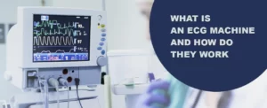
The abbreviation ECG stands for electrocardiography. The electrocardiography process entails generating an electrocardiogram, a recording of the heart’s electrical activity during repeated cardiac cycles. It is a heart electrogram that employs electrodes applied to the skin to create a graph of voltage versus time for the heart’s electrical impulses.
The test allows a doctor to pinpoint a patient’s heart rhythm and activity issues to treat their condition more effectively and precisely. The machine’s sensors, affixed to the patient’s skin, record the electrical signal generated by the heart with each beat. If the cardiologist notices any irregularities in the heart rhythm, he will advise the patient to get an ECG. When a patient experiences arrhythmias, coronary heart disease, a heart attack, heart palpitations, pain in the chest, shortness of breath, or a wildly fluctuating pulse rate, it is ideal for performing an ECG. In this blog, we’ll provide insight into electrocardiography and how it works.
Understanding the Electrocardiogram
ECG machines, also known as electrocardiogram monitors, are instruments primarily used to identify heart conditions and track the electrical activity of the cardiovascular system. It is invaluable in identifying cardiovascular conditions. These devices may determine structural abnormalities, heartbeat rate, infected or damaged tissues, surgical repairs, and more. It can figure out how certain drugs affect the heart.
Millions of cardiologists use this method, which is among the least dangerous and invasive, to find issues with the activities of the heart. The electrocardiogram is represented on paper as electrical waves. Depending on the heart’s health, these waves change.
When is an ECG used?
Doctors frequently recommend ECG in conjunction with other tests to help identify and keep track of heart conditions. It investigates heart-related symptoms like dizziness, shortness of breath, palpitations and chest pain.
An ECG can aid in identifying the following:
- Arrhythmias: A condition of abnormal heartbeat where it irregularly beats too quickly or too slowly.
- Coronary heart disease: A buildup of fatty substances can cause coronary heart disease, which causes the heart’s blood supply to be blocked or interrupted.
- Heart attacks: A condition where the heart’s blood supply is suddenly cut off.
- Cardiomyopathy: A condition in which the heart’s walls thicken or enlarge.
A person who has already been diagnosed with a heart condition or taking medication known to have potential adverse effects on the heart can also be monitored over time with a series of ECGs.
What are the types of ECG?
Electrocardiography is a crucial diagnostic tool for assessing the heart’s electrical activity. It provides valuable information about the heart’s rhythm, rate, and overall cardiac health. Several types of ECG tests serve a unique purpose in evaluating different aspects of heart function. The main types of ECG include:
Resting ECG
A resting ECG is the most common type and is typically performed in a relaxed state. Electrodes are attached to specific locations on the chest, and arms, including hands and legs, to record the heart’s electrical signals. This type of ECG helps detect arrhythmias, heart rate abnormalities, conduction disturbances, and signs of previous heart attacks. It is a simple and non-invasive procedure that provides a snapshot of the heart’s electrical activity at rest.
Stress or Exercise ECG
A stress or exercise ECG, also known as a treadmill or exercise stress test, is conducted while the patient performs physical activity, usually during a treadmill walk or a stationary bicycle ride. The purpose of this test is to monitor the heart’s response to exertion. As exercise intensity increases, the heart has to work harder, and any underlying heart conditions or abnormalities become evident. Stress ECGs help diagnose coronary artery disease, evaluate exercise capacity, and assess the effectiveness of cardiac treatments.
Ambulatory ECG
Ambulatory ECG, often called Holter monitoring, involves continuous ECG recording over an extended period, typically 24 to 48 hours. Small electrodes are attached to the chest and connected to a portable device that records the heart’s electrical activity throughout the day. This type of ECG is beneficial in capturing intermittent or elusive arrhythmias that may not be detected during a resting ECG. Ambulatory ECG comprehensively assesses the heart’s electrical behavior during normal daily activities and sleep.
Specialized ECG
In addition to these primary types, other specialized ECG tests exist for specific purposes. For instance, signal-averaged ECG focuses on detecting and analyzing subtle electrical changes associated with certain heart conditions. High-resolution ECG provides enhanced detail and resolution, aiding in detecting subtle abnormalities that may not be apparent on a standard ECG.
The different types of ECG tests serve distinct functions in assessing cardiac health. While a resting ECG provides a baseline assessment, a stress ECG evaluates the heart’s response to exercise, and an ambulatory ECG monitors electrical activity over an extended period. By utilizing these various types of ECG, healthcare professionals can comprehensively understand the heart’s electrical behaviour and make accurate diagnoses to guide appropriate treatment decisions.
How does an ECG Machine operate?
An electrocardiogram machine comprises several components that capture, amplify, and display the heart’s electrical signals. Listed here are a few of the critical elements and their roles in the functioning of an ECG machine:
Electrodes: Electrodes are small sensors affixed to specific areas of the patient’s skin. They act as receivers and transmitters of the heart’s electrical signals. A typical 12-lead ECG uses 10 electrodes, six placed on the chest and four on the limbs.
Leads: Leads are the wires that connect the electrodes to the ECG machine. They transmit the electrical signals from the electrodes to the various components within the machine. The different configurations of leads, such as the precordial leads (V1 to V6) and limb leads (I, II, III, aVR, aVL, and aVF), offer distinct perspectives of the electrical activity of the heart.
Signal Conditioning Circuitry: This circuitry within the ECG machine amplifies and filters the weak electrical signals received from the electrodes. It enhances the signal quality by eliminating noise and interference, ensuring accurate interpretation of the heart’s electrical activity.
Analog-to-Digital Converter (ADC): The ADC converts analog electrical signals into digital data that the ECG machine’s software can process. This conversion allows for further analysis and interpretation of the recorded ECG waveform.
Display and Printers: The ECG machine features a display screen that shows real-time ECG waveforms. It provides a visual representation of the heart’s electrical activity, enabling healthcare professionals to assess its rhythm, rate, and abnormalities. Additionally, the machine often includes a printer that produces hard copies of the ECG tracings for documentation and analysis.
ECG Software: The ECG machine’s software plays a crucial role in analyzing and interpreting recorded electrical signals. It uses algorithms to detect irregularities, measure intervals, and segments, and generate diagnostic reports. The software assists healthcare professionals in diagnosing various cardiac conditions, such as arrhythmias, heart attacks, and conduction abnormalities.
Power Supply: ECG machines offer flexibility for various clinical settings as either mains electricity or batteries can power them. A stable power supply ensures uninterrupted operation and precise heart electrical activity recording.
Thus, an ECG machine combines electrodes, leads, signal conditioning circuitry, ADC, display/printers, ECG software, and power supply to capture, amplify, digitise, and interpret the heart’s electrical signals. This coordinated functioning allows healthcare professionals to obtain vital information about the heart’s electrical activity, aiding in diagnosing, monitoring, and treating various cardiac conditions.
Final Thoughts
An ECG machine evaluates a heart’s rate, rhythm, and electrical activity. When a patient experiences symptoms like weakness, fainting, myocardial ischemia, heartburn, dizziness, coronary artery disease, irregular heartbeats, electrolyte imbalance, ventricular tachycardia chest pain, and many others, ECG is employed to diagnose the abnormal heart rate. The ECG monitor identifies these impulses and documents them on a graph on the monitor or paper. The heart creates electrical energy to cause it to contract. Doctors place small patched electrodes on the patient’s arms, chest, and legs during an ECG. This produces a graph, which is subsequently documented on paper. P wave, QRS complex wave, T wave, and U wave are the waves that represent the ECG readings. The graph represents the working of the patient’s heart.
Megamed takes pride in supplying hospital equipment to various healthcare institutions. We are efficient in providing various ECG machines beneficial for multiple medical practices. If the question of “ECG machine and how does it work?” arises, you can rely on the Megamed team to learn more about each ECG machine and its operations.